www.BioTopics.co.uk
Site author Richard Steane
The BioTopics website gives access to interactive resource material, developed to support the learning and teaching of Biology at a variety of levels.

Cell recognition

Cell surfaces Immune responses Vaccine/vaccination HIV/AIDS Monoclonal antibodoes ELISA test Lateral Flow Tests

All on the surface of cells

The outer surface of the cell is its cell membrane, which is a double layer of phospholipid, with a number of other substances embedded in it. These are mainly proteins, glycoproteins and glycolipids. As they project outwards into the surrounding environment, they serve as markers that identify the cell.

This topic centres on cells within the human body, and their interactions with infectious agents - mostly bacteria and viruses. Bacteria have different cell surfaces than animal cells, and the two main categories of bacteria (Gram positive and Gram negative) have distinctively different cell walls. Viruses have different outer coverings, but they are not considered to be composed of cells. In fact these superficial differences are very important.

Most biologists do not classify viruses as living organisms, although they do reproduce, but it is difficult to avoid using words like living when they are endangering the body, or killing them when they are inactivated. This distinction is also lost when discussing the production and action of vaccines.

and in the immune system

Within a multicellular organism, it is important that all the various cell types co-exist in a harmonious way. In animals, the immune system identifies and deals with any cells which are potential threats to this situation. The immune system includes white blood cells and organs and tissues of the lymph system, such as the thymus, spleen, tonsils, lymph nodes, lymph vessels, and bone marrow.

In particular, white blood cells called lymphocytes circulate in the blood and lymph and determine whether cells they encounter are self or non-self. In other words this distinction is whether they are safe and natural parts of the body (to be left alone), or foreign to the body (to be attacked and disposed of).

Lymphocytes are produced from stem cells in bone marrow, but they mature in different parts of the body. Then they make their way into the lymphatic system and circulate within the blood and in tissue fluid surrounding cells of the body.
There are two main types of lymphocytes: derived from T cells and B cells.

They respond to 'foreign' (non-self) cells by setting off an immune response.

Much of this response is controlled by receptor molecules on the cell surfaces of lymphocytes.


Note from the author

Is this topic too cumbersome?
Should it be broken up into shorter sections?


It is easy to get in touch and pass on your opinion.


T cells

Sweetbread is a culinary name for the thymus, generally from calf or lamb.
Thymus is also the scientific genus name for thyme plants.
mature in the thymus, an organ in the upper chest, above the heart and with two lobes wrapping round the trachea. Part of the maturation process involves recognition of normal ('self') cells of the body ('central tolerance'), but T cells that react to normal body cells are actively eliminated here. This ensures that that immune responses are only directed against cells of foreign ('non-self') origin. There are several classes of T-lymphocytes, which interact physically with invading organisms - the cellular (cell-mediated) immune response.

B cells

The 'B' comes not from bone but from the Bursa of Fabricius, a lymphoid organ found only in young birds
mature in the bone marrow and migrate to the spleen and lymph nodes where they are activated. They are responsible for the production of antibodies - the humoral immune response.

Both T and B lymphocytes respond to foreign antigens.

Potential threats

Users are welcome to contribute to the content below

Pathogens

All of these can enter the body and interfere with its normal functioning at the level of the cell.
Other organisms can affect the body if the immune system is in some way compromised.

Abnormal body cells

Toxins

- these are strictly chemicals, not cells.

Allergies

are conditions caused by the immune system reacting to substances as if they were harmful.

These substances - called allergens - include:
The underlying mechanism involves different cells in the body's immune system, mast cells or basophils, triggering the release of inflammatory chemicals such as histamine.

Antigens

Anything which causes the immune system to react or respond to it may be called an antigen.

When applied to cells, an antigen is a molecule of protein, glycoprotein, glycolipid or polysaccharide, on the cell surface membrane.

The outer coating of a virus can also act as an antigen. Other chemicals - perhaps not of biological origin - may also cause an immune reaction. These would then also be classed as antigens.

This results in the production (by B cells) of a specific protein called an antibody that can bind with the antigen, as a result of the two molecules fitting together due to their complementary shapes.

Adaptive immunity is protection created in response to exposure to a foreign substance.

Antigenic variability

Most pathogens have different strains or varieties, each with slightly different external proteins or other molecular components, which act as antigens.

In order to identify them, strains of disease-causing organisms are given codes based on these antigens. For example the most common current strains of human influenza virus are H1N1 and H3N2.

Memory cells will react to deal easily with pathogens of the same strain, which has antigens that have been encountered before.

However a different variety may be able to cause (worse?) infections because it has different antigens which are not recognised so quickly.

This has a number of consequences in treating these diseases and in vaccination (see below).


The different proteins, etc, which act as antigens are known as epitopes and the immune system acts on each one separately and independently.

Just the flu?

Human influenza virus spreads widely and mutates as it replicates so that different strains are produced (each year? - often named according to the perceived geographical origin), with slightly different surface antigens.

In 1918 influenza killed a large number of people - more than those killed in the 1914-1918 (first world) war. This was generally known as the Spanish flu - although it is likely that this name resulted because wartime censorship in other parts of the world prevented publicity about its effects elsewhere. It also affected more young adults than usual outbreaks, possibly because of battlefield conditions.

H stands for haemagglutinin

a glycoprotein found on the surface of influenza viruses, and responsible for their binding to cells with sialic acid on the membranes, such as cells in the upper respiratory tract.

and N stands for neuraminidase

(aka sialidase) which is an enzyme that removes the carbohydrate derivative sialic acid from mucus and glycoproteins on the surface of cells or the respiratory system. Neuraminidase (NA) on the capsid of the influenza virus particles has a role in reducing the viscosity of protective mucus, as well as attaching to host cells, and breaking out of them after infecting them.

There are 18 different haemagglutinin subtypes and 11 different neuraminidase subtypes - H1 - H18 and N1 - N11. And then there are similar influenzas in birds, pigs/swine, cattle, horses etc . . .

Cell surfaces Immune responses Vaccine/vaccination HIV/AIDS Monoclonal antibodoes ELISA test Lateral Flow Tests

The cellular response

monocyte (11K)
Monocyte
macrophage (14K)
Macrophage
White blood cells called monocytes develop into macrophages when they move into infected tissue. These are sometimes called amoeboid white cells because they can change shape and move by extending cytoplasmic processes called pseudopodia, and they surround and engulf pathogens.
These cells are called phagocytes, and the process is called phagocytosis.

Circulating pathogens (viruses or bacteria) or cells which have become infected are engulfed by macrophages. Once ingested, they are packaged into membrane-bound vesicles which interact with other vesicles (lysosomes) containing enzymes, thus inactivating or killing them.

Macrophage cells move odd molecular fragments - peptides 10-30 amino acids in length, originating from antigens of the invading foreign cells - out onto their own cell surface, where they form complexes with receptors on the cell surface and act as flags to attract other cells of the immune system.
This process is known as antigen presentation. The next few stages involve different classes of T cells.

T lymphocytes known as T-helper cells (TH cells) attach to the receptors displaying the antigen fragments on the cell surface of the macrophage and both cells release cytokines.

Interleukin-1 from the macrophage activates the T-helper cells, and they release Interleukin-2, which causes the TH cell to divide and make more TH cells, each with receptors specific to the antigen presented by the phagocyte, and also memory T cells.

These cells pass away to other parts of the immune system . . .

Cytokine also activates cytotoxic T cells (TC cells) which recognise antigen complexes on infected cells and bind to them, releasing a chemical called perforin which perforates the cell membrane of the infected cell and kills it.


More molecules on the cell surface, more acronyms

Before engulfing them, macrophages recognize pathogens by detecting specific pathogen-associated molecular patterns PAMPs on their surface. They do this via complementary pattern recognition receptor molecules (PRRs) on their own cell surface .
MHCII (108K) MHC class II receptor

After phagocytosing the pathogens, macrophages form a complex between MHC class II receptors [HLA receptors] and antigen fragments, and they move these onto the their cell surface so that they project into the intercellular fluid. MHC stands for Major Histocompatibility Complex. These MHC receptors indicate to other white blood cells that the macrophage is not a pathogen, despite having foreign antigens on its surface.

There are three types of 'professional' antigen-presenting cells, i.e. macrophages, dendritic cells, and B cells in the immune system .

Other body cells have MHC class I receptors, with slightly different protein subunits. The only exception is red blood cells (which have no nucleus). These receptors also display peptide fragments of proteins from within the cell to cytotoxic T cells, which only respond to non-self antigens.

Strange goings-on below the surface

Some infecting organisms survive after being engulfed by phagocytes and continue to live inside the protected environment of the cell. And of course they are no longer displaying their antigens, so the normal immune response is effectively prevented.

Examples include
Mycobacterium tuberculosis (causative organism of TB). This sometimes develops into granuloma tissue, and the waxy cell wall is resistant to the breakdown within lysosomes. Other macrophages engulf the infected cell so multinuclear masses are formed - these are the basic components of the tubercles.
A related organism Mycobacterium leprae (causative organism of leprosy) also forms granuloma tissue in skin and nerve tissue.
The fungus Histoplasma capsulatum can also affect the lungs. Histoplasmosis is common among AIDS patients because of their suppressed immunity.
Coxiella burnetii, the causative agent of Q fever, actually thrives and replicates in the acidic phagolysosomes of its host cell.

The humoral response

This mainly involves B cells, which go through a series of changes (activation etc). B cells are produced in the bone marrow, but they move to the spleen to mature.

Each mature B cell exhibits large numbers of receptors (50,000-100,000 or more) on its cell surface. These are basically antibodies, with the typical Y-shaped molecule (see below) attached via the stalk at the base to the cell membrane. Each receptor/antibody on a B cell is identical, and the exposed section, which has 2 antigen-binding sites, can interact with only one type of antigen, either from intact pathogens or free antigens. Many different B cells are produced, each with its own individual receptors/antibodies on the cell surface.

Pathogens attached to B cell receptors are taken in by phagocytosis and protein antigens are extracted from them so that they can be combined with receptor complexes, then displayed on the surface of the B cell - antigen presentation. This antigen is displayed alongside a large number of its antibodies.

This presented antigen is then recognized by a helper T cell specific to the same antigen, which binds to the receptor/antigen complex, and produces cytokines that stimulate the B cell to divide repeatedly (clonal expansion), producing many memory cells, each with the same receptors/antibodies on their surface (ready to repeat the process), and plasma cells which develop rough ER (with ribosomes for protein production), and these release their antibodies into the surrounding fluid.

Each B cell produces only one type of antibody. These are called monoclonal antibodies.

A foreign organism can have several different antigens on its cell surface, and the process above can be repeated so that other antigens result in extra antibodies - polyclonal antibodies.

Antibodies bind with complementary antigens on the surface of bacteria (another 'lock and key' effect), causing them to clump together - agglutination. They can also be removed by cells which perform phagocytosis.

What is a humour, anyway?

A humour is a mediaeval word used by Ancient Greek and Roman physicians and philosophers who attempted to explain the health of the body and to treat medical conditions on the basis of the balance of rather undefined factors.

The four humours of Hippocratic medicine were black bile (which gives us the expression melancholy), yellow bile, phlegm , and blood.

Of course we use the word humour to describe the contents of the eye (aqueous humour in front of the lens, and vitreous humour in the main section of the eye), but it now means simply a bodily fluid.


Polysaccharides get fast-track response

Pathogens whose cell membrane displays polysaccharides ('repeating epitopes') may bind with several sections of the antibody; this directly activates the B cell so that it proliferates into plasma cells which secrete many copies of the appropriate antibody.

Antibody structure


Antibody molecules have a characteristic Y-shaped structure, composed of 4 polypeptide chains , linked by disulphide bridges between cysteine residues.
Do not refer to these regions as 'active sites' - a term best reserved for enzymes.

At the tips of the arms are identical binding sites which interact only with a particular antigen (more technically a section of it). This section of the polypeptide chain has a distinct amino acid sequence resulting in a specific tertiary structure which gives it a certain shape (in 3-D). The shape of this site is complementary to the shape of the antigen, so that they bind together strongly to make an antigen-antibody complex.
Other antibodies have (pairs of) binding sites which differ, and consequently they will bind with other antigens. antibody (15K)
A simplified diagram of antibody structure

Each polypeptide chain has a constant (conserved) region as well as a variable region, which gives the antibody its specificity.

Within the body many different antibodies will be produced, to deal with many different foreign cell types and viruses.

There are actually a number of genes which code for the antibody polypeptides, but there is not a single gene for each type of antibody. There is a mechanism to ring the changes and make all the different proteins which make up antibodies.


Getting more globular

The geometric struts shown in some diagrams of antibodies are rather misleading.

Immunoglobulin (14K)
Antigen molecule - polypeptide chains shown in ribbon format (variable regions to left and right)
Being composed of protein, antibodies have a more fuzzy appearance due to the coiling of their polypeptide chains and the R group sidechains projecting out from the different amino acid residues which make up these chains.

And the antigen binding sites are not simply neatly curved sections, but loops forming beta sheets allowing interaction with specific sidechains.

In fact antibodies are also called immunoglobulins

There are several classes of immunoglobulins, (in addition to the thousands of different antigen specific sites). Each class has a slightly different size and anatomy which explains their roles within the body. These are known as IgM, IgG, IgD, IgE and IgA.

IgMpentamer (21K)
Pentamer of immunoglobulin M
Immunoglobulin M (IgM) is the largest class of antibody, and it is the first one to be produced (by plasma cells, in the spleen) in response to initial exposure to an antigen. The outer light chains (yellow on the right) are about 220 amino acids long, and about half of each is the 'variable region'. The longer heavy chains (blue and green) consist of about 576 amino acids. They also have variable regions composed of about 110 amino acid residues attached to 4 similar sized repeating sections and a shorter tailpiece.

Immunoglobulin M (which is itself composed of 4 polypeptide chains) tends to form groups - star-shaped pentamers or hexamers - with the antigen binding sites facing outwards.

Immunoglobulin G (IgG) is the main type of antibody found in blood circulation. It shows the typical individual Y-shaped structure.


Antibodies are actually glycoproteins, as they may have sugars (various oligosaccharides) bonded onto 5 sites (all asparagines) on the constant region of the long polypeptide chains.


What level of protein structure is shown by the antibody molecules above?
> Quaternary - 4th level of protein structure

Give an explanation for your answer
> There are several polypeptide chains or sub-units

VDJ gene rearrangement

The variation in protein structure is achieved by editing and recombining a number of component parts of the gene exons.

The gene locus encoding the H (heavy) chain variable region contains an array of about 100-300 V gene segments, about 25 D gene segments, and 6 J gene segments.

One each of the V, D, and J gene segments are selected and joined together. This is then translated at the ribosomes to make a unique polypeptide chain.

The gene locus encoding the L (light) chain variable region has two loci: κ and λ.
The κ locus consists of an array of about 40 V and 5 J gene segments.
The λ locus consists of an array of about 30 V and 4 J gene segments.

One each of the V and J gene segments are selected and joined together. This is then translated at the ribosomes to make another unique polypeptide chain.

Destruction of pathogens

antigen-antibody_complex (45K)


Antibodies bind with antigens on the outside of pathogens, forming antigen-antibody complexes.

As more and more of these stick together - agglutination - they form a semi-solid mass which settles out of the liquid in which they were suspended.

Bacterial cells with antibodies attached are also engulfed by phagocytic cells.


Opsonisation

Pathogens whose antigens have become attached to antibodies are targetted by phagocytes. These have receptors for the projecting (Fc) stalks at the base of the antibody molecule and the binding between these helps in the process of engulfing the foreign organism. An opsonin is a protein (such as an antibody or complement) that binds to foreign particles and (bacterial) cells, making them more susceptible to the action of phagocytes.

The classical complement pathway

Antigen-antibody complexes also initiate a pathway which activates the complement system (another part of the immune system, involving cascade reactions which stimulate phagocytosis and the production of cytotoxic chemicals such as the membrane attack complex).

Primary and Secondary Immune Responses


prim_sec_immune_resp (13K)
Antibody concentrations in Primary and Secondary Immune Responses


Primary response

After the first exposure to an an invading pathogen, antibodies are produced against the newly-encountered antigens on the surface of that organism.

There is a lag phase (about 1 week) where B and T cells are being activated, but not yet producing antibodies

Next, B cells differentiate into plasma cells, and secrete antibodies. At this stage fairly low quantities are normally produced (for 5-6 weeks?).

After a while, the amount of antibody decreases to minimal levels. This is due to existing plasma cells dying off, with no new plasma cells being generated to replace them. The pathogen has probably been eliminated from the body, so no further antibody production is needed.

Secondary response

On subsequent exposure to the pathogen, the immune response is much more rapid: the lag phase is typically 3-4 days. Antigen-specific memory cells quickly produce high and steady levels of antibodies.

Immunoglobulin contributions to Primary and Secondary Immune Responses

primary_and_ secondary_immune_response_IgMandIgG (37K)

In the primary immune response, antibodies produced are mostly IgM, but some IgG antibodies are also produced.

In the secondary immune response, the main antibodies secreted are IgG, sometimes with some small amounts of IgM.

Cell surfaces Immune responses Vaccine/vaccination HIV/AIDS Monoclonal antibodoes ELISA test Lateral Flow Tests

Vaccination and immunity

Vaccination is a process involving the introduction of antigens from a pathogen in order to stimulate the immune system to develop adaptive immunity to it. The material that is introduced is called a vaccine, and it is normally produced and stored under strictly controlled conditions of hygiene and scientific provenance of the source pathogen ( strain, virulence etc).

Vaccination can prevent against disease or at least reduce the seriousness of it.

Vaccination may need to be carried out several times - as 'boosters' - in order to raise the antibody level (secondary responses - see above), or if memory cells become depleted over time.

Types of vaccine

There are several sorts of material used to provide the antigens which are the active component of a vaccine:
Source material Example
'live' viruses (which are generally weaker or 'attenuated strains' of the virus) Measles, mumps, rubella (MMR combined vaccine)
inactivated or killed organisms (viruses or bacteria) Influenza
BCG Vaccine (TB vaccine)
inactivated toxins (from bacteria) Tetanus (Lockjaw)
segments of the outer covering of the pathogen (subunit, recombinant, polysaccharide, and conjugate vaccines) Human papillomavirus (HPV)
mRNA coding for segments of viral protein (spike protein) Coronavirus (Covid-19)

Sources of antigens

Bacteria can be cultured fairly easily using standard laboratory techniques: they grow on sterilised media in flasks, in incubators.

On the other hand, viruses need to grow inside living cells, so there needs to be a supply of these before they can be infected with active/'living' viruses. Live whole organisms (animals) are not used in vaccine production, but cultures of cells ('cell lines') - usually from animals, or occasionally of human origin, are often used.

There are two basic levels of supply
- master/reference cultures (originating in laboratories) - organisms/viruses proven to control the disease in question, but may have been weakened by growing at suboptimal temperature/in presence of 'attenuating agent' e.g. formalin
- production cultures - derived from the above, but not reused indefinitely, mainly for hygiene reasons.

There is a broad similarity with food or drink (cheese, yoghurt, wine, beer) production techniques which use starter cultures.

After incubation, the bacteria or viruses are extracted from the surrounding medium and processed into the form needed for the vaccine. Production of vaccines is often outsourced to a number of laboratories or vaccine plants around the world, and the techniques used need to be standardised. Quality control is problematic in some parts of the world.

In some cases preservatives are added to vaccines, and some require constant refrigeration - a problem in hot countries with intermittent power supplies.

Similar but different

The term inoculation is sometimes used alongside vaccination. In fact it is the introduction of a living organism (and I think it is OK to include viruses here) into a new environment in order for it to grow and reproduce. We inoculate bacteria onto agar to grow them in Petri dishes, or into nutrient broth in tubes. So inoculation really refers to the transfer of whole (living?) organisms, not just selected antigens from them.

Herd immunity

(community immunity)

When a sufficiently large proportion of a population has been vaccinated against a communicable disease, a situation exists whereby spread of the disease is prevented or greatly reduced. This is because even though the pathogen is released from infected individuals, there is less contact with other unprotected individuals.

This is known as herd immunity.

Some human health workers prefer the term community immunity which emphasises the commitment to public well-being, although it is rather a mouthful.

Case in point: Measles

Measles is a highly contagious virus disease. Although it has been considered to be a low-grade rash and fever, in about 1 case in 15 it may cause ear infections and neurological damage or pneumonia, and other conditions. In high income regions of the world such as Western Europe, measles causes death in about 1 in 5000 cases, but as many as 1 in 100 will die in the poorest regions of the world. Worldwide, it is still a significant cause of death.

In the UK between 1940 and 1968, notified cases of measles averaged at 400,000 per year (dipping 6 times below 200,000, and reaching 600,000 5 times), and about 100 deaths per year were reported.

After the introduction of the measles vaccine in 1968, the number of cases fell in stages, and levelled out under 20,000 from 1990 onwards. This was replaced by the MMR Measles Mumps and Rubella vaccine which gives cover against 3 viral diseases, and the NHS recommends vaccination of children at about one year old (or earlier if required), followed by a second booster (before starting school?) at about 3 years of age.

It has been stated that herd immunity is achieved with a 96% MMR vaccine coverage.

In 1998 Andrew Wakefield published a paper (now 'retracted') linking the MMR vaccine to autism and enterocolitis. By that year the number of cases of measles recorded in the UK had dropped to 56, but public reaction caused vaccination rates to fall from 90%+ to 80% in 2003/4 (and larger drops in some areas), and this resulted in a number of measles outbreaks (1370 reported cases in 2008, over 2000 in 2012).

Additionally there are a number of young people who have not had the second booster vaccination so they are not fully protected. The NHS states that children up to the age of 18 who missed, or only partially completed, their earlier MMR vaccination can have a "catch-up" MMR vaccination on the NHS.
Several large-scale investigations failed to confirm links between MMR vaccine and autism.
In 2010 Wakefield's licence to practise as a doctor was revoked by the UK General Medical Council. They found him guilty of dishonesty, the "abuse" of developmentally delayed children by giving them unnecessary and invasive medical procedures, and acting without ethical approval for his research. He has moved to America and continues to campaign on autism.


A similar situation has occurred in other parts of the world, but this has been exacerbated by global travel and pockets of unvaccinated people of different ethnic groups.
See web links below

There are controversial anti-vaccination campaigns which feature prominently on social media ....

Active and passive immunity

Active immunity is immunological protection against foreign organisms as a consequence of exposure to them or their antigens, as explained above. The production of antibodies takes some time to take effect, but once established it lasts for a long time - all life long in some cases.

Passive immunity is the transfer of antibodies produced in another organism and its effect is generally short-lived.

Mothers and babies

Antibodies which have developed (over time) in the blood of a pregnant female can be passed across the placenta to the blood of the developing foetus so that it has acquired a certain amount of immunity to pathogens it may encounter after birth.

Antibodies can also possibly be passed via breast milk to the new-born baby. Colostrum, the thicker initial secretion from the mammary glands, contains a number of ingredients that assist development of new-born babies.

Antivenoms against snake bites etc

If an animal e.g. a horse or a sheep is injected with (small, but then increased) doses of snake venom, it will develop antibodies against it. Antibodies can be extracted from the blood and purified for use as a treatment for humans bitten by the same or similar species of snake. This may be called antivenin or venom antiserum.

A similar situation exists with scorpion stings, spider bites and stings from certain fishes.


Small pocks - big success

Note the past tense
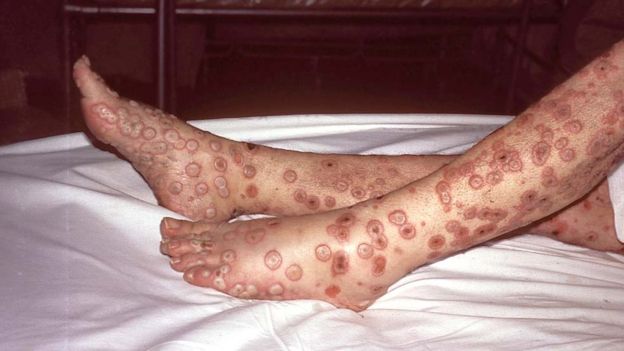
Smallpox was a very contagious disease caused by a virus - often called variola. Symptoms included fever followed by rash on the tongue, spreading to the face and all over the body, with sunken sores ('pocks') containing an opaque fluid, eventually forming scabs which fell off. It is said that it was fatal in 30% of cases, and easily passed from person to person. It was known about three thousand years ago, spreading along trade routes between Africa, Asia and Europe, reaching the Americas in the sixteenth century.

Variolation was a rather random technique that involved giving someone a limited exposure to the virus, through material from scabs of infected people being sniffed up into the nose, or placed under the cut skin. This caused a less severe form of the smallpox (which could still spread and cause an epidemic), and usually gave immunity to those so treated. However, about 1-2% of treated people died (which is better than 30%).

By 1700, variolation was fairly commonplace in Africa, India and the Ottoman Empire, and it was introduced to England in the 1720's.

Some key names

Edward Jenner (1749-1823)

was a doctor in Berkeley, Gloucestershire. Apparently he underwent variolation as a schoolchild, and that impaired his health somewhat. As a doctor, he performed variolation on his patients. Living in the country, he knew about cowpox, a disease of cows, showing as pocks on the udder, and he heard that people who caught it from their cows could not subsequently catch smallpox.

Sarah Nelmes

was a milkmaid who had a rash on her hand, which Jenner diagnosed as cowpox.

Blossom

was the cow who passed it on to Sarah as she was being milked (by hand).

James Phipps

was the eight-year old son of Jenner's gardener, and Jenner transferred fluid from one of Sarah's pocks to 'scratches' on James' skin. He became poorly but quickly recovered. One and a half months later Jenner subjected James to variolation with smallpox material, but he did not become infected and remained immune to smallpox for the rest of his life.

Jenner published his results and went on to develop his findings and supply cowpox material (which was in fairly short supply). He encountered opposition from other medics and other sceptical people, but he was honoured and recompensed in a number of ways.

Vaccinia

is the name given to cowpox, based on the Latin vacca for cow. This gives its name to vaccination.

The use of cowpox vaccine gradually received acceptability to the public. In fact variolation was forbidden by Act of Parliament in 1840 and vaccination with cowpox was made compulsory in 1853.

Janet Parker

was the last person to die of smallpox, in 1978, astonishingly in the UK.
She was a medical photographer at the Birmingham University Medical School and she worked one floor above the Medical Microbiology Department where smallpox research was being conducted. As a consequence, work on smallpox around the world was dramatically scaled down.

Finishing off this disease

Following vaccination campaigns, smallpox was considered to be eliminated in North America in 1952 and Europe 1953, leaving South America, Asia, and Africa. In 1959 the World Health Organization (WHO) launched a campaign to eradicate smallpox worldwide but they had to re-launch it in 1980. At that time they estimated that there were still up to 15 million cases of smallpox each year. Teams of vaccinators from all over the world journeyed to the remotest of communities to vaccinate every person in the areas at risk. In 1980 it was officially declared that smallpox had been eradicated. It is the only worldwide disease that has been eliminated so far.

All stocks of smallpox virus have been destroyed, with the exception of material kept under high security in the USA (CDC, Atlanta) and Russia (State Research Center of Virology and Biotechnology) in Koltsovo.

Less of a pest

In 2010, the Food and Agriculture Organization announced that the viral disease rinderpest (in the same group as measles), which infected cattle and other ruminants had been completely eradicated, by the use of a live attenuated vaccine, making this the first (and so far the only) disease of livestock to have been eradicated.
measles_global_coverage_2017 (126K)

More candidates

Measles and rubella are seen as possible candidates for eradication by continued vaccination programmes.

The risk of polio has been greatly reduced in many parts of the world. Its incidence has decreased by 99.9 percent; from an estimated 350,000 cases in 1988 to a low of 483 cases in 2001, after which it remained at a level of about 1,000 - 2000 cases per year for a number of years. In 2015, cases decreased to 98 and further decreased in 2016 to 37 wild cases and 5 circulating vaccine-derived cases, but increased in 2018 to 33 wild cases and 103 circulating vaccine-derived cases.

Polio is currently the subject of a global eradication program. Another is Dracunculiasis, also called Guinea-worm disease, a parasitic infection by the Guinea worm.


Thicker than water?

In the ABO series of blood groups, there are 4 types of cells. There are 3 possible different antigens on the surfaces of red blood cells, and 3 possible antibodies in the blood plasma.

Blood group A B AB O
Antigen(s) A
B A and B "none"
- but see below
Antibodies anti-B anti-A none anti-A and anti-B
Antigen oligosaccharide chains
on red blood cell membranes


key (1K)


aantigen (2K)

bantigen (2K)
Both of these antigens are found on each AB red blood cell's membrane
aantigen (2K)


bantigen (2K)


hantigen (2K)

The basic (precursor) oligosaccharide chain

Genes and enzymes involved

I stands for isohaemagglutinogen

The A allele (IA) encodes 1-3-N-acetyl galactosaminyl transferase that bonds α-N-acetyl galactosamine to the D-galactose end of the H antigen

The B allele (IB) encodes 1-3-galactosyl transferase that bonds α-D-galactose to the D-galactose end of the H antigen

The O allele (IO or i) contains a nucleotide deletion that results in a loss of enzymatic activity

Cell surfaces Immune responses Vaccine/vaccination HIV/AIDS Monoclonal antibodoes ELISA test Lateral Flow Tests

HIV

Structure of the human immunodeficiency virus (HIV)

HIV1 (34K)
Image credit: National Institute of Allergy and Infectious Diseases National Institutes of Health


In the centre of the HIV particle is its genetic material, which is RNA. This is unlike most living organisms - all eukaryotic and prokaryotic organisms and lots of viruses - which use DNA.

There are also some rather unusual enzymes which allow the RNA to take over a cell of the human body.

In a mature virus particle, this is enclosed inside a layer called a capsid, made of many round protein units joined together in rings to form a bullet-shaped protective layer.

On the outside of this is a much more uniform and spherical covering (envelope) consisting of a layer of phospholipid taken from the membrane of the cell in which the virus developed.

Projecting from this and evenly spaced around it are a number of spikes consisting of glycoproteins (gp120 and gp41 above). These function as attachment proteins and allow the particle to target, bind to and infect T helper cells (TH) or macrophages which have the appropriate receptors on their surface.

Some arty images of HIV

Count the 'spikes'! HIVarty (265K)
Out of proportion ? HIV+RBC (198K) Red blood cells (7.5 µm) are about 60 times larger than HIV particles (120nm)


There are two versions of HIV - HIV1 and HIV2 (mostly confined to western Africa).
These are not each shown above.
They are called lentiviruses because they are slow growing.

More detail

The outside of the HIV virion is spherical, and covered with a phospholipid bilayer, derived from the (previous) host cell as it budded off when leaving.

Attached to this are about 72 'spikes' - virus-produced glycoproteins (gp120 and gp41), with a carbohydrate section (glycans, mostly built up from mannose units) shielding the underlying protein. Each of these is present as a trimer - a group of three. Gp120 functions as a docking protein - binding with CD4 receptors on cells (which also have CCR5 and CXCR4 co-receptors).

Immediately inside this is a layer of (P17) protein units forming a supportive matrix.

The main or central part of the virus particle is bounded by a capsid made of p24 protein (2000 subunits) with a curious tapering cone-like or bullet shape.

Inside this are:
the viral genome - 2 sections of single-stranded RNA - partly covered by protein ('nucleocapsid')
and a few enzymes: reverse transcriptase, integrase,
plus 4 proteins which act as control factors in taking over the host cell's metabolism.

Replication of HIV in helper T cells


replicationCycleHIV (43K)
Image credit: National Institute of Allergy and Infectious Diseases National Institutes of Health


The HIV virion attaches to a target T cell, by its external spikes binding to receptors and co-receptors on the cell surface, and the cell membrane and outer viral envelope fuse.

The central part of the HIV virion enters the cell.

The capsid is broken down and the (single stranded) RNA is released into the cell's cytoplasm, as 'genomic RNA'. It is accompanied by with a special enzyme reverse transcriptase, which converts it into a short section of (double stranded) DNA.

Put simply, in this process, inside the cell:
- a single-stranded length of HIV DNA is made from the RNA.
- a complementary strand to the HIV DNA is made, in the same way as a new complementary strand is made during semi-conservative replication of human DNA

You should be able to describe how the complementary strand of HIV DNA is made.
> (Complementary) nucleotides/bases pair (A to T and C to G)
> You might get credit for these being drawn together by hydrogen bonding
> This is DNA polymerase activity
> Nucleotides join together (to form the new strand) - phosphodiester bonds form

You should also be able to say how this copying process differs from the normal replication of DNA.
>No need for strands to separate/unzip
>No helicase enzyme involved

This 'proviral' DNA (effectively of viral origin) is then inserted using another viral enzyme integrase into the cell's own DNA, within chromosomes inside the nucleus.

This integrated DNA can remain within the cell for quite a long time and it is shielded from surveillance by the immune system. Such cells can be considered as a latent reservoir of the virus. There are medical strategies to attack actively replicating stages in the virus cycle but the latent reservoir is more difficult to deal with.

At a later stage, this section of DNA can function like a normal gene in the host cell. It can undergo transcription, and produce mRNA, as well as copies of RNA (genomic RNA) like the one that infected the cell in the first place.

The mRNA can use the cell's normal protein production process (ribosomes, tRNA etc) to make more of the viral enzymes and control proteins. The proteins are produced in a row, like a string of sausages, which is then broken down into single proteins by a viral enzyme protease.

Virally-coded proteins and RNA accumulate near the edge of the cell, and become enclosed inside a newly-formed capsid shell. This buds out of the cell, taking a section of the cell membrane to form its envelope.

It is estimated that 1000-3000 virus particles are released from each infected cell. These travel round in the blood stream, and attack any more T cells or macrophages that they encounter.
Target cells are mostly T helper cells (TH) or macrophages or dendritic cells, all of which have CD4 receptors on their surface, so they are called CD4+ cells.

Attachment to these causes the gp120 trimers to open like a flower and CD4 and gp41 to change in shape so that the HIV virion also binds to a coreceptor CCR5 or CXCR4 and the central part of the HIV virion then enters the cell.

HIV belongs to a class of viruses called retroviruses. They are called this because they perform a process that is the reverse of the usual stage of transcription (DNA → (m)RNA) [which is then translated to form protein].

Retroviruses are RNA (ribonucleic acid) viruses, and in order to replicate (duplicate) they must make a DNA (deoxyribonucleic acid) copy of their RNA.

This is achieved using the enzyme reverse transcriptase.

The processes by which viral DNA is produced and integrated into chromosomes are not as reliable as normal DNA copying in the cell (which is subject to its own 'proofreading' process). This undoubtedly contributes to the high mutation rate characteristic of HIV, which results in many different varieties or strains of the virus.

The reverse transcription process

Reverse transcriptase does not simply start at one end of the single strand of HIV RNA and move along converting it into 2 strands of DNA. In fact there are 3 distinct types of activity.

HIV RNA → DNA


RNA in red, DNA in Blue

revtransaction (47K)


A
It starts at a primer binding site (PBS) only about a third of the way back from the 5' end of the RNA. A tRNAlys3 molecule is shown attaching here.
B
A complementary copy is made from RNA to single stranded DNA (RNA-dependent DNA polymerase activity):
(G to C, C to G, A to T, U to A).
This copying stops at the 5' end of the RNA strand, which is composed of R (repeat section). It is accompanied by the breakdown of the RNA which has been 'copied' (ribonuclease H activity).
C
The DNA strand then 'jumps'to the other end of the HIV RNA which also has a R region, so the end of the short section of single stranded DNA anneals with the RNA.
D
More ss DNA is made, until the 5' end of the RNA, and most of the RNA is again broken down. Only a middle section PPT (polypurine tract) is left.
E
Formation of the second DNA strand proceeds fron PPT using the other ssDNA as a template (DNA-dependent DNA polymerase activity):
(G to C, C to G, A to T, T to A).
F
Copying ends at the PBS section (and the remaining RNA is broken down).
G
There are now 2 incomplete strands, overlapping at the PBS section
H
Each DNA strand rebuilds itself starting from the 3' ends, using the other strand as a template.
There is now a LTR (long terminal repeat) at each end of the dsDNA

Integrase action

Integration of HIV proviral DNA into host cell DNA integration (15K) Click on image above for animation
(Mouseover returns to first frame)
The HIV DNA has the base sequence CAGT on both strands on the 3' end.

Integrase cuts off 2 bases, leaving an exposed section on the 5' ends. This is is similar to the 'sticky ends' produced by bacterial restriction enzymes as used in recombinant DNA to transfer plasmids.

Integrase - which appears to act in groups of two or four, also cuts the host cell DNA but leaves staggered ends 5 base pairs long. When the viral DNA inserts itself, there is a gap 3 bases long on each strand. This appears to be filled in by host cell's enzymes.

How HIV causes the symptoms of AIDS

HIV is spread most commonly during sexual intercourse, heterosexual or homosexual.
It enters via mucosal linings, and other sexually transmitted infections can increase the risk of viral infection by damaging these tissues.

Children can gain infection from their mother before or during birth. The virus may also be transmitted from an HIV-infected mother to her infant by breast-feeding.

HIV also can be transmitted by contact with infected blood. This is linked with drug taking and the sharing of needles or syringes contaminated with minute quantities of blood containing the virus.

In the past, blood transfusions have spread the infection, but this is very rare nowadays as blood and products are routinely screeened.

Primary HIV infection

Most people infected by HIV develop a flu-like illness (primary or acute HIV infection) within 1-2 months after the virus enters the body. (Show/hide more detail:)

HIV does not directly kill the infected person but it does weaken the immune system, so diseases caused by other opportunistic pathogenic organisms or cancers can become established because of the lack of T cells (and macrophages). Untreated, HIV typically turns into AIDS in about 10 years. This is when the number of T cells in the body has declined to very low levels so the immune system cannot resist infections.

The relationship between HIV viral load - red line - and TH lymphocyte counts - blue line - over the average course of untreated HIV infection
Hiv-timecourse (57K)
Image credit:Wikipedia


Progression to AIDS

Acquired immunodeficiency syndrome (AIDS) is a chronic, potentially life-threatening condition caused by the human immunodeficiency virus (HIV).
(Show/hide more detail:)
Opportunistic infections may occur as a result
(Show/hide more detail:)
In HIV/AIDS certain cancers are common
(Show/hide more detail:)

Anti-HIV strategies

Enzyme inhibitors

Compounds inhibitory to three enzymes originating from the HIV virus particle can be potential candidates for interrupting the infection cycle. These are sometimes referred to as highly active antiretroviral therapy (HAART) treatments, and they involve regular and continuous administration of drugs, usually in tablet form. They are sometimes made available as combination medications.

Reverse transcriptase inhibitors

could prevent the viral RNA from producing DNA.
Examples include
Tenofovir - a modified version of d-AMP which causes termination of the DNA strand, and emtricitabine - a cytidine analogue. Both of these are included in the drug branded as Truvada, and other generic brands.
Efavirenz acts allosterically by binding to a distinct site away from the active site of the reverse transcriptase enzyme, known as the NNRTI pocket.
AZTjs (2K) Azidothymidine (AZT), also known as Zidovudine (ZDV) is a thymidine analogue, causing termination of the DNA chain as it forms from viral RNA. This has been widely used medically to prevent mother-to-child spread during birth or after other potential exposure such as a needlestick injury.
[ Cautionary note: In a clinical trial conducted by National Institute of Health for the drug Fialuridine, a thymidine analogue having antiviral activity against Hepatitis B virus, showed adverse reactions in phase 2 clinical trials leading to death of five human volunteers due to severe hepatic toxicity and lactic acidosis. This was found to be due to production of defective mitochondrial DNA resulting in high levels of lactic acid and deposition of fat in mitochondrial microvesicles.]

All of the compounds mentioned above have their 3-D molecular structure revealed in files on this website: see links below.

Integrase inhibitors

could prevent DNA from being incorporated into chromosomes inside the T cell nucleus. Raltegravir is an example of this.

Both of these strategies (inhibiting reverse transcriptase and inhibiting integrase) could be used

Protease inhibitors

prevent the release of proteins required for new virus particles. Many versions are available, all with names ending in -avir.

Early versions acted as peptidomimetics (which mimic a peptide linkage- particularly between phenylalanine and proline or tyrosine and proline), found to be critical sequences in the polypeptide chain joining distinct sections of the virally-coded polypeptide chain.

A number of other molecular approaches have also been approved, and factors such as the solubility and bioavailabilty of different compounds also enter into consideration.

The enzyme HIV protease has been well studied and details of its amino acid composition and catalytic action are well known. It is a dimer of 2 units, each with 99 amino acid residues. In particular, two aspartic acid residues have a key role in binding substrate in its active site

Vaccines

The HIV virus's external glycoproteins gp120 seem like ideal candidates for vaccine production, but the mannan section on their outside does not cause a clear antigen-antibody reaction. So there is not a current anti-HIV vaccine. It is thought that a number of people have developed immunity to HIV and some of these are under examination to establish the details of this.

Targetting co-receptors

Alongside the CD4 receptors that bind with HIV glycoproteins are other cofactors: CCR5 and CXCR4. It has been found that some people lack these due to a change in the gene coding for this protein. Direct gene therapy to mimic this is impracticable, but it has been found that compounds called chemokines produced by CD8+ cells can block access to these co-receptors.

Stem cell transplantation

It has recently been discovered that individuals being treated for cancer (lymphoma) by transplantation of stem cells from another individual after radiation treatment to kill cells in the bone marrow have acquired immunity to HIV as a consequence of the stem cell donor lacking the gene making CCR5. This is very fortunate, but it is unlikely to be repeated routinely.

Why antibiotics are ineffective against viruses

An antibiotic is a chemical substance produced by a bacterium or fungus, which kills or prevents growth of another species of microorganism (usually a bacterium, possibly a fungus). Alternatively it may be produced by another organism. Earthworms, poison dart frogs and tropical plants are other odd possible sources. Some people do not use the term antibiotic for purely chemical antimicrobial compounds of synthetic origin.

The effectiveness of antibiotics rests on the specificity of their action against their target microorganisms - principally those causing diseases of Man (or animals). They must act against a feature that is only found in the pathogenic microorganism, not in the human or other 'patient'. For example, penicillin causes the production of bacterial cell walls to fail, resulting in bacterial death due to osmotic damage. Other antibiotics, e.g. erythromycin, kill bacteria by preventing bacterial protein synthesis.

The key feature is that antibiotics are much more toxic to bacteria than they are to human cells.

You should be able to say why these antibiotics do not affect humans
penicillin > Humans do not have cell walls (membrane is outer layer of cells)
erythromycin > Humans use a different type of ribosome for protein synthesis.
Bacterial ribosomes are smaller than human ribosomes (20 nm against 25-35 nm in diameter, or 70S vs 80S sedimentation coefficient)

By the same token, viruses are not affected by antibiotics because they do not have cell walls or any organelles, and they are not metabolically active.

But viruses can be affected by antibodies produced by B cells in the human body.

Cell surfaces Immune responses Vaccine/vaccination HIV/AIDS Monoclonal antibodoes ELISA test Lateral Flow Tests

Monoclonal antibodies

Monoclonal antibodies (MABs for short) can be used to seek out cells in the body which have a particular antigen on their surface, or to react to an antigen in bodily fluid.
Many medical conditions are caused by cells with different antigens on their surface.

I have put in the trade names of well-known brands of these mAb products in a medical context, together with the scientific names (all of which end -ab).

Antigen-antibody reactions against cancer

Cancer cells may self-destruct (by apoptosis) when antibodies attach to them

Cancer cells coated with antibodies are more likely to be sought out by cells of the immune system and removed by phagocytosis.

Monoclonal antibodies may be used to interfere with some of the stages in cancer development: vascularisation (growth of blood vessels which supply nutrients), or the prevention of cell messaging by blocking cancer cell growth promoters, as well as immune system inhibitors.

For example
Avastin (bevacizumab) is an mAb that targets a protein called VEGF (vascular endothelial growth factor) that affects tumour blood vessel growth.
more/ less detail

Herceptin (Trastuzumab) is a drug that recognises HER2 (human epidermal growth factor receptor 2) on cancer cells.
more/ less detail

Humira - the name derived from "human monoclonal antibody in rheumatoid arthritis" - (Adalimumab) is a medication used to treat rheumatoid arthritis and a number of related conditions. It does this by binding to tumor necrosis factor-alpha (TNFα), so it has an anti-inflammatory action. more/ less detail

Targeting medication to specific cell types

This can be done by attaching a chemotherapeutic drug to a monoclonal antibody.
These are called conjugated monoclonal antibodies.
In this way the treatment is delivered directly to the unwanted cells whilst avoiding healthy cells. This would be an example of medicine or drug acting as a 'magic bullet' or a 'silver bullet'.

For example
Kadcyla (ado-trastuzumab emtansine) targets the HER2 protein which is over-expressed in some breast cancers, attached to a chemo drug called DM1 - mertansine/emtansine which is a tubulin inhibitor and it inhibits the assembly of microtubules by binding to tubulin.

Radioimmunotherapy relies on the targeted delivery of radiation to kill cancer cells.

For example
Zevalin (Ibritumomab tiuxetan) is a radiolabelled mAb i.e. a monoclonal antibody against the CD20 antigen, which is found on B cells (lymphocytes). It can be used to treat some types of non-Hodgkin lymphoma.

Medical diagnosis

Some conditions are characterised by the production of specific proteins which serve as markers for that condition, and monoclonal antibodies can be used in detecting these antigens.

For example:
pregtest (99K) Pregnancy test kits use monoclonal antibodies.
Antibodies to a hormone called HCG - human chorionic gonadotrophin - which is found in the urine of pregnant women, are used in pregnancy test sticks.
If she is pregnant, HCG in a woman's urine will bind to the monoclonal antibodies on the test stick, and this will cause a change in colour or pattern which will indicate pregnancy. This is a form of lateral flow test. See below


Prostate specific antigen (PSA) is a marker for prostate cancer. (See ELISA test below)
more/ less detail

Carcinoembryonic antigen (CEA) is produced normally in embryonic development, but it is also found in elevated concentrations in the blood as a result of colorectal and related cancers.
more/ less detail

Testing for microbial infections

HIV
more/ less detail

Coronavirus Covid 19

See Lateral Flow Tests below


Polyclonal and monoclonal


When a person (or a lab animal) becomes infected with a pathogenic organism, antibodies will be produced in response to the antigens on the surface of that organism. There may be several types of antibody, if there are several antigens on the outside of the organism. Each antibody is produced by a single B cell, which will divide to make more identical antibody-producing cells (a clone) within the body (each producing the same antibody). As there are usually several antigens on the outside of the infecting organism, it is quite normal to end up with a mixture of polyclonal antibodies to its antigens and this is quite efficient in dealing with pathogenic organisms.

Similarly, chemical compounds which are not normally found in the body can be injected, and antibodies to them will be produced, usually by a single set of B cells.

Within the blood stream will be a number of antibodies, not just the ones mentioned above. They can be extracted, purified and separated, but this is quite a tedious process.

In the laboratory it is often useful to have a pure sample of antibodies to a single antigen or chemical - monoclonal antibodies.

Production of monoclonal antibodies

This can be achieved by removing B cells from the spleen of a lab animal (usually a mouse) which was previously exposed to the antigen in question. However, once outside of the body these cells would soon die, so they are fused with myeloma cells from a culture line derived from a tumour (which continues to grow, even outside the body).

The resulting hybridoma cells can be cultured in flasks etc, and a single type of antibody can easily be extracted from the liquid media. In fact it is necessary to separate individual cells and screen them to see what antibody they are producing. But once this has been done, the cell can be encouraged to grow and produce more cells, each producing the same monoclonal antibody.

There is another issue: useful antibodies produced in one species of mammal can be recognised as foreign by another species so they would be rejected by the immune system. They need to be 'humanised' by the replacement of some of the immunoglobulin polypeptide chain with the human equivalent.

A more universal alternative - in the future

(Thousands of) genes can be extracted from human cells and incorporated into microbial cells by 'standard' recombinant technology, resulting in a library of many different antibody-producing cells. This will involve very large scale screening, but avoid problems with the humanising process..

A personal perpective

2018 Chemistry Nobel Prize winner, Sir Gregory Winter Life Scientific radio programme

Sir Gregory Winter
Gregory Winter (15K)
Program blurb:
In an astonishing story of a scientific discovery, Greg Winter tells Jim Al-Khalili how decades of curiosity-driven research led to a revolution in medicine. Forced to temporarily abandon his work in the lab when a road rage incident left him with a paralysed right arm, Greg Winter spent several months looking at the structure of proteins. Looking at the stunning computer graphics made the pain in his arm go away. It also led him to a Nobel Prize winning idea: to ‘humanise’ mouse antibodies. A visit to an old lady in hospital made Greg determined to put his research to good use. He fought hard to ensure open access to the technology he invented and set up a start up company to encourage the development of therapeutic drugs. It took years to persuade anyone to fund his Nobel Prize winning idea that led to the creation of an entirely new class of drugs, known as monoclonal antibodies. In 2018, the market for these drugs, which include Humira for rheumatoid arthritis and Herceptin for breast cancer, was worth $70 billion.

Ethical issues


Production of vaccines involves killing animals to provide populations of cells in which viruses are cultured. Monkey kidney cells have been widely used, as well as duck embryo cells and dog kidney cells. Live chicken's eggs have been used for culturing viruses.

There are also two main populations of cells of human origin, taken (in the 1960s) from aborted foetuses which are widely used in vaccine production. A cell strain known as WI-38 was developed by Leonard Hayflick working at the Wistar Institute, using lung cells from an aborted foetus. Similarly a cell strain MRC-5 originating from foetal lung cells has been developed at the Medical Research Center in the United Kingdom. These cells can be frozen in liquid nitrogen and revived for use in vaccine production but they are not 'immortal' in the sense of hybridomas obtained by combining 'ordinary' cells with cancer cells. It is important to note that these cell strains do not rely on continued supply of material from abortions.

The rubella vaccine uses viruses isolated from a foetus which was terminated at the request of a pregnant woman after getting the infection.

The use of material originating from aborted foetuses is unacceptable to many, not just Roman Catholics. More recently cells derived from surplus embryos produced by IVF have been used for research, but they are still considered morally questionable by many.

In various parts of the world, especially in Africa and Asia, vaccines have been suspected to have completely different effects than the protection expected. This may be caused by mistrust of the West and America in particular. In particular it has been suggested that the purpose behind vaccination programmes is to sterilise women. The effects of these rumours has been documented in Cameroon and Tanzania in the 1990s, and it also adversely affected the uptake of measles vaccination in Nigeria in 2005. Other suspicions have been that vaccines in fact spread AIDS or cancer. In Pakistan and Afghanistan, vaccination has been objected to on religious and political grounds by Muslim fundamentalists . Attacks on polio vaccination teams have also occurred, thereby hampering international efforts to eradicate polio in Pakistan.

The production of monoclonal antibodies involves the use of live mice, which are killed when their spleens are removed to get B cells.

The use of transgenic mice, which have been given human genes, raises questions over the ethics of genetic engineering.

Testing of drugs and vaccines on primates (macaques, Chimpanzees etc) - the nearest relatives to Man - is unacceptable to some, and they may not necessarily be the most dependable test organisms.

Testing of potential drugs and vaccines on Man carries risks - and some object to 'blind trials'

Scare stories

Monoclonal over-reaction

In 2006, a monoclonal antibody Theralizumab (also known as TGN1412), intended for use against certain forms of leukaemia and arthritis was tested on six (paid) human volunteers, alongside two who were given a placebo.

The drug was administered to all the participants in a short period of time, and all six receiving the substance under test soon felt distress, headache, fever and pain. They were all transferred to a local hospital intensive care unit and given corticosteroids to reduce inflammation, and plasma-exchange to attempt to remove TGN1412 from their circulation. Four suffered from multiple organ dysfunction. One person lost fingers and toes as a result. They all left hospital 1-3 months later but their immune systems may be somewhat compromised

The condition that caused these symptoms is now known to be cytokine release syndrome, with marked swelling of skin and mucous membranes, as in a severe allergic reaction.

The antibody was produced to target a T-cell co-receptor CD28 and it was thought that it could be therapeutically useful in stimulating the immune system in immunosuppressed patients.

Although the dose admistered was thought to a low safe starting point, it was in fact later found that a near-maximum immuno-stimulatory dose had been given and it would bind to 86 to 91% of all CD28 receptors in the body,

It was tested on cynomolgus monkeys (crab-eating macaques) Macaca fascicularis. It was initially thought that, being raised in the laboratory, they had not been exposed to infections and so did not have memory T cells which were stimulated by the antibody. It was later found that these monkeys lack CD28 expression so their effector T-cells could not be stimulated by the drug.

Polio vaccines

Two distinctly different approaches have competed for precedence in worldwide initiatives to control poliomyelitis. This is a virus disease affecting several parts of the body, but infection of nervous tissues is generally characterised by paralysis of the legs.

Jonas Salk's polio vaccine (inactivated with formalin) came into use in 1955. This was given by injection, and sometimes caused mild redness or pain at the site of injection.

Albert Sabin's oral polio vaccine (a mixture of attenuated live strains, with well-defined mutations following passage of the virus through nonhuman cells at a lower temperature than human body temperature) came into commercial use in 1961. This is said to revert or mutate back very occasionally, causing about three cases of vaccine-associated paralytic poliomyelitis per million doses given, which is considered negligible in light of casualties caused by polio epidemics. It was recommended by the US National Institutes of Health, but some in countries there is a trend towards inactivated polio vaccine.

The Cutter incident

In 1955 it was found that 250 patients in the US contracted paralytic polio about a week after being vaccinated with Salk polio vaccine from Cutter pharmaceutical company, and the paralysis was limited to the limb the vaccine was injected into. Some lots of Salk polio vaccine had not been properly inactivated, allowing live poliovirus into more than 100,000 doses of vaccine.

Polio vaccine problem in Maharashtra 2018

India was officially declared "polio free" by the government in March 2014, following intensive vaccination programmes.
However it was found that some vials of the oral polio vaccines contaminated with type-2 polio virus (said to be eradicated worldwide) were administered to children in Maharashtra and Telengana.

Foot and mouth - containment or contamination?

Foot and mouth is a serious and contagious virus disease of cattle, but it rarely affects humans. An outbreak in Shropshire UK in October 1967 was traced to remains of legally imported infected lamb from Argentina and Chile. The virus spread and a total of 442,000 animals were slaughtered (estimated cost £370 million).
In 2001 there was an epidemic that resulted in the slaughter of around ten million sheep and cattle, costing Britain £8 billion. This was traced to infected meat that had been illegally imported to Britain. It was caused by the "Type O pan Asia" strain of the disease.
In August 2007 foot-and-mouth disease was discovered in Normandy, Surrey, UK. The strain of the virus was identified as a "01 BFS67-like" virus, one linked to vaccines and not normally found in animals. It was in fact the same strain as the 1967 outbreak.

This strain was used at the nearby Institute for Animal Health (specialising in virus research) and Merial Animal Health Ltd (vaccine production) at Pirbright, 2.5 miles away. Though technically separate establishments, they were found to have shared leaking waste pipes and inadequate hygiene procedures. The premises were identified as a probable source of infection but no prosecution took place because inspectors were unable to pinpoint the exact source of the outbreak.

In November 2012 and January 2013, there were breaches in biosecurity when a ventilation system was operated 'in a different configuration from normal', at the Pirbright Institute, for which it was fined £72,350.

Cell surfaces Immune responses Vaccine/vaccination HIV/AIDS Monoclonal antibodoes ELISA test Lateral Flow Tests

The ELISA test

ELISAplate (196K)
ELISA stands for enzyme-linked immunosorbent assay.

It is used to measure the concentration of biologically important chemicals in liquids.

The test involves monoclonal antibodies to the test substance which have been chemically combined with an enzyme and immobilised by attachment to a surface. Often two separate monoclonal antibodies to the same test substance are used. The number of antigen-antibody complexes formed is in direct proportion to the concentration of antigens in the sample.

After the antibody-enzyme complex has had a chance to react with the antigen, the excess liquid is poured away, and the substrate to the enzyme is added and incubated.

This results in the release of a coloured chemical. The concentration of this is also in direct proportion to the concentration of antigens in the sample. The depth of colour can be read using a spectrophotometer and the optical density used to calculate the concentration of the antigen,

It is often performed in multi-welled polystyrene plates and is often used in high throughput laboratories.

There are several versions of the test: the most common are direct, indirect and sandwich.

Confirming mumps with ELISA

Mumps is a viral disease which is easily spread in university/college scenarios.
It is characterised by swelling of salivary glands, headache, fever, and sometimes inflammation of the testes.

A test has been developed to find out whether a person has antibodies against the mumps virus.

The test shown in the diagram below (not to scale) is an example of the indirect ELISA technique.

ELISAmumps (59K)
Why will this test detect mumps antibodies, but not other antibodies to other viruses such as measles or rubella in the blood?
>The first antibodies are specific to the (trapping) mumps antigen - because they have the appropriate shape/ tertiary structure, and the second antibodies are also specific to the first - mumps - antibody.
Antibodies to other viruses will not bind in step 2.


Why is it important to wash the well at the start of Step 4?
>This removes unbound second antibodies (with enzyme attached)
Otherwise enzyme may still be present/you may get (stronger) colour change anyway - false positive.


Why will there be no colour change if mumps antibodies are not present in the blood?
>There will be no antibodies to bind to the antigen, so the second antibody (with the enzyme) will not bind either.
So no enzyme/enzyme-carrying antibody will be present (after washing in step 4).


Enzymes and Substrates used

Enzyme Substrate Colour Absorption wavelength
alkaline phosphatase p-nitro­phenyl phosphate yellow 405 nm
(Horse­radish) peroxidase o-phenyl­diamine dihydro­chloride orange-brown 450 nm
β-galacto­sidase o-nitro­phenyl-beta-D-galacto­pyranoside (ONPG) yellow 410nm


Detecting PSA with ELISA

An antigen called PSA is present in the blood of men in the early stages of prostate cancer.

There is a blood test for PSA. The test uses monoclonal antibodies to PSA. The stages in the test are shown in the diagram.

This is an example of the sandwich ELISA technique, which relies on two different monoclonal antibodies to (two separate epitopes on) the target PSA antigen.


PSAELISA (34K)


In the second stage, PSA binds to some of the bound antibody.

What would cause the PSA to bind to some, but not all, of the antibody?

> less PSA than bound antibodies/so test is within acceptable range of detection, i.e. reliable

If PSA bound to all of the bound antibody at this stage, what would that signify?
> There could be more PSA than bound antibodies/test result may be above level of discrimination, so may not be reliable/needs dilution for proper titration

If the final stage did not result in a coloured product, what would be the conclusion?
> No PSA in the blood sample - all clear!

Cell surfaces Immune responses Vaccine/vaccination HIV/AIDS Monoclonal antibodoes ELISA test Lateral Flow Tests

Lateral Flow Tests

also known as Rapid Antigen Tests

COVID-19 Self-Tests

These have become very widely used as a consequence of the Covid-19 pandemic, for testing people without the classical symptoms of infection by the virus, and in the NHS Test and Trace process.

Lateral flow tests detect proteins in the outer covering of the virus particles, in contrast to PCR tests, which detect the RNA inside the coronavirus.
PCR samples must be sent to a specialist laboratory for testing following the reverse transcriptase-polymerase chain reaction (RT-PCR), which obviously takes some time to process, as well as being more expensive.

Nasal swabs are used to transfer virus particles into a liquid, 4 drops of which is simply placed into a test cassette, and results are achieved with 15-30 minutes.

The process is called lateral flow testing because it relies on the liquid spreading out sideways on a flat surface so that any antigen in it can interact with monoclonal antibodies and be carried along the strip enclosed in it. This is similar to the sandwich ELISA technique (although no enzymes are involved), but it uses coloured lines to show the result.

A coloured line in the T region confirms the presence of (antigens from) Coronavrus CoV-19, but there must also be a line in the C region to confirm the validity of the test.

Possible results

COVID-19-Antigen-Rapid-Test-Kit-Result-InterpretationJOYSBIO (78K)

Principle of operation

The swabs are agitated in a liquid - also called a buffer solution - containing a surfactant molecule which penetrates membranes, presumably releasing proteins from the viral capsid.

Much has been published about the spike proteins which are involved in viral attachment to target cells, and their variation in different mutants, but there is also a nucleocapsid (N) protein. This has a larger molecule, and there are several positions (epitopes) where different antibodies can become attached.

The sample pad, beneath the nozzle ('specimen well') through which buffer sample is introduced, allows the liquid to flow sideways across the 'conjugation pad'. This contains one monoclonal antibody to the coronavirus N protein, labelled with colloidal gold nanoparticles, and so the protein (if present) can bind and form an antigen-antibody complex with it. This complex is carried across to an area where another monoclonal antibody to N protein is tethered to the film, in a straight line. This can also bind with the labelled antigen-antibody complex, resulting in a noticeable red-coloured line across the strip, in the test ('T') line region.

The conjugation pad also contains an unrelated protein immunoglobulin Y - Ig Y - similarly labelled with gold particles by means of a different labelled monoclonal antibody, and this is carried across the test line without binding to the antibody there. It continues to the second, control ('C') line where another monoclonal antibody is also tethered. The appearance of a coloured line here is taken as confirmation that the test procedure itself has been successful, with the correct sample size and flow across the membrane. Otherwise it is classed as a void or invalid result.

Lateral flow in action


triangle (1K) LateralFlowAnimation (97K)

Thanks to the animals

Nucleoprotein antibody is initially obtained from rabbits, yolk cells from chicken eggs provide the Ig Y and the monoclonal antibody developed to it involves goats.

Cell surfaces Immune responses Vaccine/vaccination HIV/AIDS Monoclonal antibodoes ELISA test Lateral Flow Tests

Other related topics on this site

(also accessible from the drop-down menu above)

Similar level

Transport across cell membranes
Eukaryotic cells
Virus Particles

Simpler level

Antigens and antibodies - The antigen-antibody reaction, development of monoclonal antibodies
Blood - components and their functions
Viral diseases - The main ones affecting humans, signs and symptoms, control/prevention, means of spread

Interactive 3-D molecular graphic models on this site

(also accessible from the drop-down menu above)

The 3-dimensional structure of Human Chorionic Gonadotrophin - I have chosen to use hCG to illustrate the four levels of protein structure

3-D molecular structure of AZT - The azide group at C3' puts a stop to the developing proviral DNA chain from HIV
3-D molecular structure of Tenofovir - More of a nucleotide, but completely lacking a 3' carbon
3-D molecular structure of emtricitabine - Sulphur , not carbon at the 3' position, so no phosphodiester backbone
3-D molecular structure of Efavirenz - Not a nucleoside, but an allosteric inhibitor of RT
3-D molecular structure of Fialuridine - A nucleoside, but with fatal consequences: Is it the C2' substitution, the iodine, or the arabinose?

Web references

The Adaptive Humoral Immune Response - nice animation

B Lymphocytes and Humoral Immunity from Lumen Learning

The Humoral Immune Response 6:12 Youtube video animation from the Cancer Research Institute [nothing new from 4:14]

Differences between Primary and Secondary Immune Response - from Microbiology Notes: "All notes of Bacteriology, Virology, Parasitology, Mycology and Laboratory"

Influenza 101 virology blog

Types of Influenza Viruses Centers for Disease Control and Prevention

About Edward Jenner - from The Jenner Institute

Smallpox - from Centers for Disease Control and Prevention

How smallpox claimed its final victim By Monica Rimmer BBC News

The History of Vaccines from The College of Physicians of Philadelphia

RETRACTED: Ileal-lymphoid-nodular Hyperplasia, Non-specific Colitis, and Pervasive Developmental Disorder in Children Andrew J. Wakefield et al. The Lancet 637-41 (1998)

Measles, misinformation, and risk: personal belief exemptions and the MMR vaccine from the Journal of Law and the Biosciences

Measles: Half a million UK children missed vaccine - reaction from the BBC to Unicef report

New York county declares measles outbreak emergency - more from the BBC

Measles Cases and Outbreaks - US CDC updates (Centre for Disease Control and Prevention)

Measles Key facts from the World Health Organisation (WHO)

Measles eradicated in UK in 'sign of recovery' after MMR vaccine controversy - a (slightly over-optimistic?) report from The Independent - and what does that large image of cells portray?

HIV life cycle: How HIV infects a cell and replicates itself using reverse transcriptase - YouTube video

HIV/AIDS From Wikipedia, the free encyclopedia

How HIV Causes AIDS National Institute of Allergy and Infectious Diseases National Institutes of Health [archived version]

HIV-1 Ribonuclease H: Structure, Catalytic Mechanism and Inhibitors by Greg L. Beilhartz and Matthias Götte

UK patient 'free' of HIV after stem cell treatment BBC News story

Discovery and development of HIV-protease inhibitors From Wikipedia, the free encyclopedia

Co-Receptors: CCR5 -- Understanding HIV This web page bears the following warning:
This article is part of The Body PRO's archive. Because it contains information that may no longer be accurate, this article should only be considered a historical document.
However, it predicted the potential of stem cell transplantation.


PrEP (pre-exposure prophylaxis) - Terrence Higgins Trust

Adalimumab From Wikipedia, the free encyclopedia

Bevacizumab (Avastin) From Wikipedia, the free encyclopedia

Trastuzumab emtansine (Kadcyla) From Wikipedia, the free encyclopedia

Monoclonal antibodies to treat cancer - from the American Cancer Society

Three immunoassays based on monoclonal antibodies specific for prostate specific antigen (PSA), alpha-1-antichymotrypsin (ACT), and the PSA-ACT complex.

Diagnosis of HIV/AIDS From Wikipedia, the free encyclopedia

Theralizumab From Wikipedia, the free encyclopedia - an alarming story of adverse test reactions to TGN1412 an immunomodulatory monoclonal antibody drug

Pirbright Institute fined over foot-and-mouth breach

After contaminated vaccine sparks polio fears in India, WHO calms nerves By Kuwar Singh [polio vaccine problem in Maharashtra 2018]

A New Vaccine for Tuberculosis: The Challenges of Development and Deployment

ELISA - technical stuff

Raltegravir: molecular basis of its mechanism of action

JOYSBIO Coronavirus Antigen Rapid Test Kit

In silico discovery of antigenic proteins and epitopes of SARS-CoV-2 for the development of a vaccine or a diagnostic approach for COVID-19- a 'study aimed to discover likely antigenic proteins and epitopes to be used for the development of a vaccine or serodiagnostic assay using an in silico approach'

Coronavirus epitopes predictions (from the above) showing amino acid sequences (8-20+) of 9 or 10 'probable antigen' nucleocapsid epitopes

www.BioTopics.co.uk    Home     Contents     Contact via form     Contact via email     Howlers     Books     WWWlinks     Terms of use     Privacy