Eukaryotes are normal higher animals and plants, and fungi, as well as
protoctists, algae,
as distinct from prokaryotes (bacteria, cyanobacteria and archaea).
In fact the term Eukaryota is used as a taxonomic group (Domain) above Kingdom.
Their cells contain a true nucleus, and several membrane-bound structures (organelles).
These membranes each consist of a double layer (a 'phospholipid bilayer'), with varying amounts of protein and other substances embedded in it.
Mouseover text beneath pictures for more information
Click on pictures for enlarged version (opens in separate window)
ANIMAL CELLS
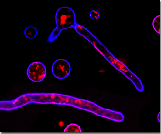
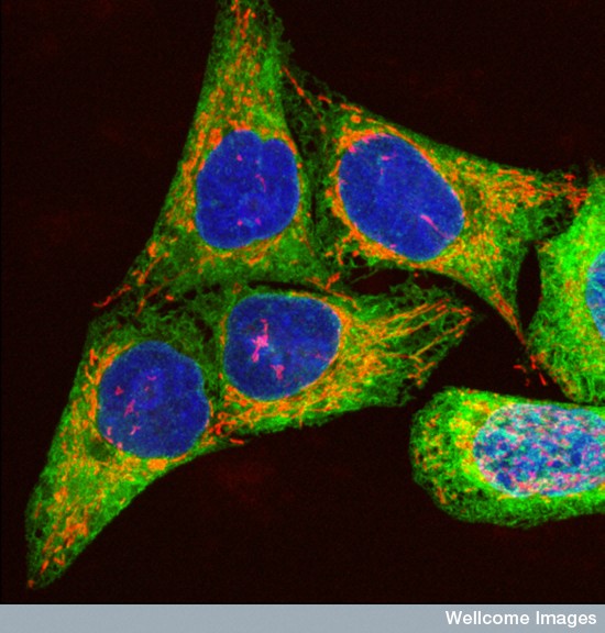 Human melanoma cells showing DNA, mitochondria and ER
- Paul J.Smith & Rachel Errington, Wellcome Images
Human melanoma cells showing DNA, mitochondria and ER
- Paul J.Smith & Rachel Errington, Wellcome Images
GREEN PLANT CELLS
Nuclei are often hard to see in plant cells, as they are hidden by other components.
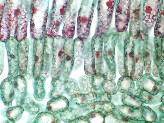 Mesophyll cells from the lilac Syringa
- University of Wisconsin
Mesophyll cells from the lilac Syringa
- University of Wisconsin
ALGAL CELLS
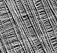
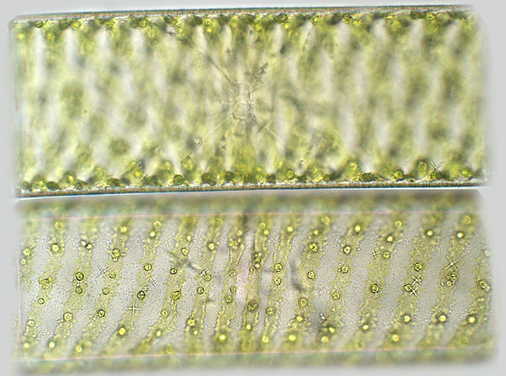 Spirogyra
 - a filamentous alga
- University of Wisconsin
YEAST CELLS
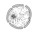
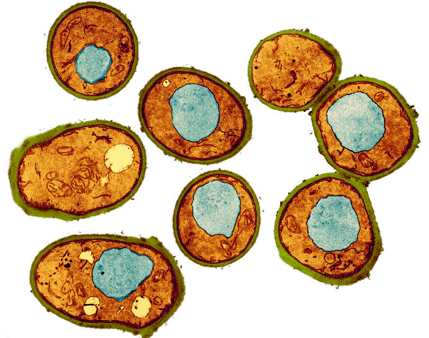 Yeast cells, coloured transmission electron micrograph (TEM)
- Thomas Deerinck, Ncmir
Yeast cells, coloured transmission electron micrograph (TEM)
- Thomas Deerinck, Ncmir
FUNGAL HYPHAE
 Sections of hyphae of Trichoderma reesei
- Mari Valkonen, VTT Finland
Sections of hyphae of Trichoderma reesei
- Mari Valkonen, VTT Finland
FUNGAL HYPHAE
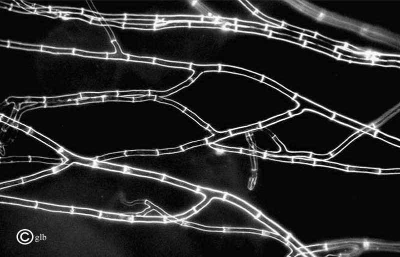 Septate hyphae of of Sordaria fimicola - not really cells
- from George Barron's Website on Fungi
Septate hyphae of of Sordaria fimicola - not really cells
- from George Barron's Website on Fungi
Cell specialisation
In a multicellular organism, different cells become specialised to perform different functions.
This is often reflected in a changed number of these features,
e.g. increased numbers of mitochondria to produce more ATP for extra activity - in liver (1000-2000 mitochondria per cell), muscle etc, or more ribosomes where protein synthesis occurs.
Sometimes specialised cells have unique features such as the actin and myosin filaments of muscle cells, or haemoglobin in red blood cells, which also lose their nucleus as they develop.
Levels of organisation
(cells, tissues, organs, systems, individuals)
When a number of similar
cells work together, they are called a
tissue:
tissues are generally named the same as the individual cells: e.g. muscle is built up from muscle cells.
Several different tissues perform slightly different tasks contributing to the overall function of an
organ:
muscle tissue, nervous tissue and lining tissue all contribute to the functioning of the heart;
muscle tissue, nervous tissue and lining tissue together with secretory tissue all contribute to the functioning of the stomach.
Organs are combined to form different
systems, e.g.
heart, arteries and veins form the circulatory system (arguably in conjunction with all the other organs of the body),
the digestive system consists of the alimentary canal with all its specialised regions which are organs in their own right, e.g stomach, ileum, colon, and associated glands such as salivary glands, liver and pancreas.
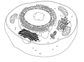
Cell components seen in transverse section
(mouseover for labels)
Eukaryotic cells contain the following components:
 Nucleus
Nucleus,
surrounded by a double membrane, with pores. Within this are a number of chromosomes: each one a single, long linear DNA molecule, wound onto basic (alkaline) histone proteins, organising the DNA.
Nucleosomes are formed from about 150 DNA base pairs wrapped round a molecular spool formed from eight histone subunits, and
the DNA continues to wind round many other nucleosomes.
This DNA-protein complex is called
chromatin, a reflection of its ability to absorb stain.
Euchromatin is fairly diffuse - allowing access to enzymes responsible for transcription during normal day-to-day operation of the (non-dividing) cell.
Heterochromatin is more dense and tightly packed, so it stains more darkly.
Normally, chromosomes are not distinctly visible as individual thread-like structures until the cell starts to divide.
Within the nucleus one or more
nucleoli can sometimes be seen. These sometimes look like featureless blobs. They are the site of ribosomal RNA synthesis, alongside certain chromosomes.
There is normally only one nucleus per cell. Only cells about to divide have two nuclei.
Nuclei in eukaryotes duplicate by the process of mitosis, before the cell itself divides. This requires the cell to re-organise itself so as to separate replicated chromosomes into two groups using a structure called the spindle, composed of microtubules.
Plasma membrane
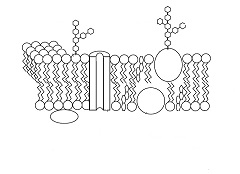
Cell surface-membrane (plasma membrane) a flexible phospholipid bilayer, with different proteins - channel and carrier molecules passing from one side to another, other proteins on the inner or the outer surface and various substances such as glycoproteins and glycolipids embedded in it but projecting into the extracellular space. Although this layer is elastic and appears to give the cell its shape, it is not under pressure like a balloon and its structure is always forming and re-forming. This
fluid mosaic is stabilised by hydrophobic interactions between phospholipid fatty acid tails on the inside of the bilayer and hydrophilic interactions between the phospholipid heads which face out into the extracellular fluid on one side and into the cytoplasm on the other.
 Rough endoplasmic reticulum
Endoplasmic reticulum, a network of interlinked flattened sacs called cisternae. In sectional diagrams and electron microscope photographs they may look like tubes.
Smooth endoplasmic reticulum is composed of standard membrane - phospholipid bilayer - and is associated with the production and metabolism of lipids and steroid hormones.
Rough endoplasmic reticulum has a granular appearance due to ribosomes attached to the outside of the membrane. It is involved in protein production: translation, folding of the polypeptide chain to give the protein its tertiary structure, as well as sending the resulting protein onwards in vesicles.
Ribosomes are the site of protein synthesis, and are composed of 2 RNA subunits, together with proteins.
In eukaryotic cells they are a distinctive size: 25-35 nm in diameter, but they are usually described as
80S, with subunits 60S and 40S (Svedberg sedimentation coefficients). Some mammalian cells contain several million ribosomes.
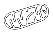
A mitochondrion
Mitochondria, cylindrical or ovoid structures with an inner and an outer membrane, the inner one folded into shelf-like cristae to give more surface area.
Aerobic respiration takes place in mitochondria. The tricarboxylic acid cycle takes place in the matrix inside the inner membrane, and oxidative phosphorylation (producing ATP) on the inner face of the inner membrane. Metabolically active cells will have more mitochondria. Liver cells typically contain 1000-2000 mitochondria. Heart muscle cells probably have more. Sperm cells have many in the middle region, which leads into the flagellum. Cells carrying out active transport always have prominent mitochondria.
Mitochondria have their own DNA (mtDNA) and ribosomes, smaller than normal eukaryotic ribosomes. This acts as confirmation for the
endosymbiont theory which suggests that these organelles represent an independent organism taken into the cell and then incorporated into it.
In most species including humans, mtDNA is inherited solely from the mother, via the fertilised egg. The sperm loses its mitochondria before fertilisation.
It shows less variation than other genetic markers and is widely used in comparing populations, in evolutionary biology and phylogenetics.
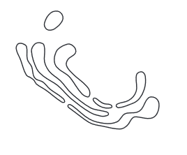 Golgi apparatus
Golgi apparatus - also known as Golgi body - a membranous structure rather like a stack of plates or dishes, with membrane-bound vesicles budding off from its edges.
Lysosomes are one type of Golgi vesicle which store
lysosomal enzymes - a variety of hydrolytic enzymes which become active when a lysosome fuses with another organelle, the internal pH falling to about 5.0.
In some cells, secretory lysosomes are actively involved in the storage and delivery of the cell's specific products. Conventional lysosomes are involved in the dismantling and re-cycling of various substrates presented to them within the cell. In particular they are responsible for returning amino acids to the cell for re-use. Several hundred lysosomes may be present in a single animal cell.
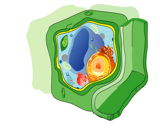
In addition to these components, plants and algae have the following features:
 Cellulose fibres in cell wall
Cell wall on the outside of the cell, (with the cell membrane usually pressed hard against it).
This is fairly firm and gives a distinct shape to these cells.
The cell wall is normally made of a basket of cellulose fibres so water-soluble substances can easily pass through it.
Fungi have a cell wall but it is generally composed of other polysaccharides, e.g. chitin, not starch.
One or more
vacuoles - fluid-filled spaces within the cytoplasm - effectively lined by a membrane - the tonoplast. Vacuoles may vary in size in response to osmotic pressure, but usually occupy 30-90% of a cell by volume.
Plant cells are normally turgid as a result of hydrostatic pressure in the vacuole, acting against the cell wall. This is responsible for the rigidity of the plant as a whole.
In early stages of cell growth, whilst the cell wall is in the process of formation, this pressure can result in cell elongation.
Vacuoles also store soluble chemicals, including nutrients.
They also function to break down complex molecules. Proton pump molecules in the vacuolar membrane (similar to those in mitochondria and chloroplasts, but using up ATP rather than generating it) pump in H
+ ions, maintaining acid conditions for hydrolase enzymes which break down un-needed chemicals.
Vacuoles also act as stores for particular chemicals produced by different parts of the plant: organs of perennation (rhizomes, tubers, corms and bulbs) and seeds contain food stores - proteins, oils and carbohydrates. Some of these chemicals can be quite distinctive: pigments in petals, odoriferous defence chemicals such as those produced by the onion family, latex in a variety of plants.
Some of these are extracted for use as drugs: digitalis from foxgloves, opium from poppies.
Fungi also have (smaller) vacuoles.
Plants and algae are likely to have the following features in some of their cells (in leaf palisade layer and spongy mesophyll, also in guard cells, and occasionally in other green parts, e.g. stems):
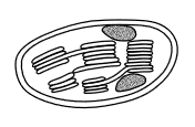 A chloroplast
Chloroplasts, green ovoid structures with
three membranes: both an outer and an inner membrane round the outside, and also a number of inner membranous structures consisting of interleaving sheets called thylakoids, containing chlorophyll. These enclose a space or lumen, and some sections of the thylakoid are stacked on top of one another forming grana. Photosynthesis takes place in chloroplasts: the light-dependent reaction in the grana, and the light-independent reaction in the fluid filled spaces on the outside of the thylakoids (stroma).
Like mitochondria, chloroplasts have their own DNA and ribosomes, smaller than normal eukaryotic ribosomes. This acts as confirmation for the
endosymbiont theory which suggests that these organelles represent an independent organism taken into the cell and then incorporated into it.
Other cell components
Centrioles
Cytoskeleton
Peroxisomes

 Home
Home